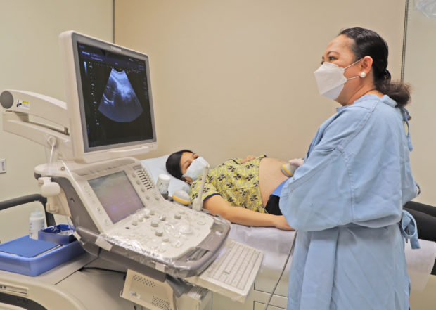
Multidisciplinary treatment advances in unborn babies’ heartbeat irregularities are saving lives
One of the routines in pregnancy care is screening for congenital abnormalities. While most babies are born without birth defects, there is always a possibility that a risk of abnormal development will present itself regardless of a mother’s or father’s age, lifestyle, or family or personal history.
Congenital anomaly screening is one tool to detect structurally critical and significant congenital heart defects prenatally. When you’re pregnant, you only want the best for your unborn baby. Thus, a high-risk pregnancy or unexpected diagnosis can be really scary.
Such is the case of a 26-year-old lady with twin pregnancy who was diagnosed at 22 weeks age of gestation with supraventricular tachycardia (SVT) and hydrops fetalis in one of the twins.

Regular monitoring of pregnancy helps the health care provider identify potential health problems early and take steps to manage them to protect the health of both mother and the unborn child.
Fetal arrhythmia is an abnormal or irregular heartbeat pattern that can occur while the baby is still in the womb. Some types of arrhythmias are relatively benign and will not affect the baby at all while others can be life-threatening. Fetal SVT is the most frequently reported type of fetal arrhythmia which refers to a rapid heart rate rhythm that begins in the upper chambers of the heart. Most cases are benign and transient and do not require treatment. However, persistent fetal tachycardia, if left untreated, may lead to fetal hydrops, a serious, life-threatening condition characterized by excessive fluid in the different parts of the body of the fetus.
The case report “Transplacental therapy of supraventricular tachycardia and hydrops fetalis in a twin pregnancy” highlights that while the management of multifetal gestation affected by SVT can be challenging due to scarcity of information, with early diagnosis, successful multidisciplinary treatment and strict monitoring, good fetal and neonatal outcomes can be achieved.
Authored by Dr. Joseph Carl Mariano Macalintal and Dr. Maria Rosario Castillo-Cheng, Perinatologists from the Institute for Women’s Health, The Medical City (TMC) Ortigas, and Dr. Dexter Eugene D. Cheng, Pediatric Cardiologist and Interventionalist from the same institution, the case report has been presented and published both locally and internationally. This unique and interesting case report has won first place and was commended in the recent Philippine Society of Maternal Fetal Medicine Fellows’ Forum and was published in the International Journal of Prenatal Cardiology.
This medical literary publication reports on a patient pregnant with twins who went for a congenital anomaly scan in another institution and was incidentally found to have on her 22nd week of pregnancy a problem in one of her babies. One twin had fetal tachycardia with its heart rate registering at 242 bpm (normal fetal heart rate is 120–160 beats per minute). The other sonologic findings on Twin A included skin and scalp edema and pleural effusion (an unusual amount of fluid in the lungs).
The patient was referred to Dr. Castillo-Cheng who recommended that a fetal 2D echocardiography be done at The Medical City Ortigas. The fetal 2D echocardiogram, a useful ancillary tool in the diagnosis and management of fetal arrhythmia was conducted by Dr. Dexter Eugene Cheng. Through this fetal echo it was determined that Twin A was suffering from cardiogenic hydrops fetalis secondary to supraventricular tachycardia. The second of twin, Twin B, was found to be normal and without any cardiac abnormalities. The patient was then advised admission at The Medical City Ortigas for further work-up and management of Twin A’s condition.
Despite the condition of Twin A, immediate delivery was not a plausible option because of the extreme prematurity of both twins. The multidisciplinary team, along with full consent obtained from the patient and her husband, opted to administer medical transplacental antiarrhythmic treatment.
“Our plan was to closely monitor the patient and her twins while on medical treatment, push the pregnancy as close to term as possible and deliver the babies at 37 weeks age of gestation so that we could avoid the complications associated with prematurity,” says Dr. Castillo-Cheng.
Twin A significantly improved with the initiation of medical treatment. Its hydropic state resolved without its sibling, Twin B and their mother manifesting any deterioration nor untoward severe complications. Good growth was achieved and an unremarkable antenatal course ensued thereafter. The patient was able to reach term and delivered by cesarean section at 37 weeks due to fetal breech presentation. The babies were born with good Apgar scores. Their heart rates were both normal and with regular rhythms. Although the electrocardiogram (ECG) at birth of Twin A who was earlier diagnosed with SVT showed normal sinus rhythm, his chest X-ray showed cardiomegaly (enlarged heart), which Pediatric Cardiologist Dr. Dexter Eugene Cheng managed accordingly. Twin A was discharged on his seventh day of life after the completion of his medications and a 2D echocardiography that showed normal cardiac structure and function.
Dr. Castillo – Cheng says that aside from prematurity that made immediate delivery not possible, there were other factors that made this case challenging and interesting. For one, the management of SVT in multifetal pregnancies, cases of which are even less frequently encountered, is not yet defined unlike in singleton pregnancies.
“Several case reports and studies have been performed and reported abroad regarding both the success and failure of transplacental anti-arrhythmic therapy in singleton pregnancies, but information on the successful management of multifetal gestation affected by fetal tachycardia is scarce to none,” adds Dr. Castillo-Cheng.
The uniqueness of this case is primarily the management of the isolated arrhythmia in one of the twins made more complicated by the possible adverse effect of the anti-arrhythmic medications on the unaffected twin and on the mother as well.
“Dr. Dexter Cheng and I had to consider the potential risks that may occur with the antiarrhythmic therapy to the mother and the other normal fetus , the ability to provide adequate monitoring and the mother’s willingness to submit to such therapy,” says Dr. Castillo-Cheng.
She further explains that once a decision to treat was made, close fetal and maternal follow-up was required to detect and manage side effects earlier. A multidisciplinary team conference attended by the Maternal Fetal Medicine team and the Pediatric Cardiology service led by Dr. Castillo – Cheng and Dr. Dexter Eugene Cheng respectively was held to discuss with the patient and her family the possible adverse effects of the medications and the plans for close fetal surveillance which included a biweekly fetal growth and cardiac monitoring. Regular monitoring of complete blood count, serum electrolytes, and liver enzymes were also done during the pregnancy.
As the case report has cited, “the simultaneous administration of Digoxin and Amiodarone may successfully treat atrial fibrillation and induce cardioversion in the affected twin with careful monitoring for untoward side effects allowing the prolongation of pregnancy, and thereby avoiding the complications of preterm delivery .”
“It was a happy and successful outcome for this pregnancy. The hydrops was completely reversed in Twin A and postnatal evaluation of the twins did not show any abnormalities,” says Dr. Castillo-Cheng. Despite being so, the parents were advised of the possible course of Twin A’s condition, the possibility of recurrence in later life, and the importance of long-term follow-up.
To read the full case report, visit: https://www.termedia.pl/Transplacental-therapy-of-supraventricular-tachycardia-and-hydrops-fetalis-in-a-twin-pregnancy-case-report-and-literature-review,146,45065,1,1.html
For inquiries on The Medical City’s maternity services, please send an email to [email protected].
ADVT