A better test that detects hidden cardiac ‘time bomb’
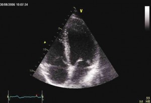
You probably know what an echo is—sound waves that are bounced off after hitting a substance or structure. That’s exactly what an echocardiogram is: a test using a device that emits high-frequency sound waves directed across the chest. The resulting echo is then picked up by the same device, which is then converted into images that could be viewed on-screen.
This test gives a detailed view of the structures of one’s heart, helping doctors determine how well (or how poorly) a particular heart is working.
“Interestingly, this test should be widely utilized more than chest X-ray or the more popular electrocardiogram (ECG), which measures and records the electrical activity of the heart,” says Dr. Adolfo Bellosillo, president and founder of the Foundation for Lay Education on Heart Diseases (FLEHD), a nonprofit organization advocating proper heart care and heart disease education.
Bellosillo, who is also the head of the Makati Medical Center’s Cardiac Rehabilitation and Preventive Cardiology Unit, said one can no longer just rely on ECG results as they could be inaccurate and are prone to erroneous interpretation.
An ECG is widely used because it is able to screen for a variety of cardiac abnormalities. Moreover, ECG machines are readily available in most medical facilities, and the test is simple to perform, risk-free and inexpensive.
Useful test
“However, a number of individuals who are predisposed to sudden cardiac death may be given a clean bill of health based on a flawed ECG interpretation. On the other hand, taking an X-ray of the chest is not always reliable as it could only show the external border of a heart chamber and not the inner border, which is important in detecting if an abnormal growth or thickening of heart muscle is present.
Presence of a thickened heart muscle may be a potentially lethal disease. This overdeveloped heart muscle is less efficient at pumping blood throughout the body and may also affect the electrical activity of the heart, which can lead to abnormal heart rhythms or even sudden death.
This is why Bellosillo often reminds resident physicians and cardiovascular fellows-in-training to be careful in immediately discharging a patient who came to the emergency room complaining of chest discomfort but whose ECG result showed “normal” findings.
“The ECG can often be normal or nearly normal in patients with undiagnosed coronary artery disease or other forms of heart disease (this is called false negative results) while at the same time, many ‘abnormalities’ that appear on the ECG result turns out to have no medical significance after a thorough evaluation is done (false positive results),” he says.
However, if one adds echocardiogram, Bellosillo explains that the doctor will now have a more reliable means in visualizing cardiac structures, cardiac walls (especially presence of abnormal thickening) and even the velocity of blood flow at certain points in the heart.
More judicious
Bellosillo says: “For decades ECG and chest X-ray were relied on heavily because there were no better methods or alternatives. With the availability of echocardiogram, I believe doctors should now be more judicious in relying on ECGs or even chest X-rays. Yes, an echocardiogram test may be more expensive, but it can be very valuable, particularly in identifying whether there is already damage to the heart muscle or tell the extent of heart muscle damage if one had just suffered a heart attack.”
Painless and does not use radiation, echocardiogram only requires the patient to lie on a couch, turned on his/her left side. The doctor will use a device that is moved across the chest (a clear gel is rubbed over the chest to help the device slide on skin). This same probe that emits high-frequency sound detects the same high-frequency sound waves that have bounced back, creating a moving image of the heart on a screen.
The whole procedure normally takes between 15 and 45 minutes.

